Saturday 28 September 2013

Biofilm And Plaque --Precursors to Dental Diseases

The Diseases of the gums can broadly be classified into
  • Gingivitis
  • Periodontitis
GINGIVITIS is defined as an inflammation of the GINGIVA only.When the inflammation progresses to the other parts of the PERIODONTIUM it is termed as PERIODONTITIS.

Before we go into the nitty gritty world of gingivitis let,s understand the beginning of gum disease.
Many of you will be wondering that this is not possible as I take care of my teeth to the nth degree, but my friends don't forget about the bad bacteria lurking in your mouth!!!

As soon as we brush our teeth squeaky clean we remove any growth or covering on our tooth surfaces.
But this does not remain so as our teeth are constantly under a wet environment. The first step in plaque biofilm development is the 
adsorption of host and bacterial molecules to the tooth surface.

 Within minutes of tooth eruption or a cleaning, PELLICLE 

formation begins, which can be defined as a thin coat of salivary 

proteins. The pellicle acts like an adhesive 

by sticking to the tooth surface and encouraging a conditioning film

 of bacteria to attach to the pellicle. This 

conditioning film directly influences the initial microbial 

colonization, and continues to adsorb bacteria to the

 tooth surface.This early composition of the biofilm is able to 

withstand many of the frequent mechanisms of the oral cavity that

 contribute to bacterial removal such as swallowing, nose blowing, 

chewing, and salivary 

fluid outflow. The early colonizers are also able to survive in the 

high oxygen concentrations present in the 

oral cavity, without having much protection from other bacteria .

 Thus, this thin, initial biofilm is almost always 

present on the tooth surface as it forms immediately after cleaning. 

Passive Transport of Oral Bacteria to the Tooth Surface


Following pellicle formation, there is passive transport of oral 

bacteria to the tooth surface, which involves a reversible adhesion

 process. By using weak, long-range physicochemical interactions

 between the pellicle coated tooth surface and the microbial cell

 surface, an area of weak attraction is formed that encourages the 

microbes to reverse their previous adhesion to the pellicle and come 

off the tooth surface (hence the term "reversible adhesion"). This 

reversible adhesion then leads to a much stronger, irreversible 

attachment, as short-range interactions between specific molecules

 on the bacterial cells and the complementary receptor proteins on

 the pellicle surface occur. Because many oral microbial species

 have multiple adhesion types on their cell surface, they can thus

 participate in a plethora of interactions with both other microbes 

and with the host surface molecules.


Co-Adhesion of later colonizers to already attached early colonizers


The co-adhesion of the later colonizers to the already
 present biofilm continues to involve many specific 
interactions between bacterial receptors and adhesions.
 These interactions build up the biofilm to create a more
 diverse environment, which includes the development of
 unusual morphological structures like corn-cobs and 
rosettes.
The many interactions between these diverse bacterial 
species begin to create a number of synergistic and 
antagonistic biochemical interactions. For example, bacterial 
residing in food chains may help to contribute metabolically 
with other bacteria if they are located physically close to one
 another. Similarly, when obligate anaerobes and aerobes 
are involved in co-adhesion, these interactions can ensure 
the anaerobic bacteria’s survival in the oxygen-rich oral 
cavity.

Multiplication of the Attached Microorganisms


Eventually, the bacterial cells continue to divide until a three-

dimensional mixed-culture biofilm forms that is spacially and 

functionally organized. Polymer production causes the

 development of the extracellular matrix, which consists of

 soluble and insoluble glucans, fructans, and

 heteropolymers. This matrix is one of the key structural

 aspects of the plaque biofilm, much like that of other

 biofilms. Biofilms such as this are very thick, consisting of

 100-300 cell layers. The bacterial stratification is arranged

 according to metabolism and aerotolerance, with the

 number of gram-negative cocci, rods and filaments

 increasing as more anaerobic bacteria appear. As the

 biofilm thickens and becomes more mature, these anaerobic 
bacteria can live deeper within the biofilm, to further protect 

them from the oxygen-rich environment within the oral 

cavity.This biofilm is now composed of a variety of bacteria 

and is now known as DENTAL PLAQUE.

Plaque formation


At 24 hours the maturing dental plaque contains a wide variety of bacteria and it is possible to detect easily identifiable inter-species associations such as the well documented "corn-cob-configurations", although a wide variety of other inter-species associations will be present.
Further colonisation and growth of established bacteria takes place as the plaque matures to form a stable, climax, community. This pattern of development leading to a climax community has been termed "bacterial succession". The resulting community consists of individual microbes and microcolonies acting in complex consortia which can convey a range of beneficial properties. These include feeding synergies, improved antibiotic resistance and a host of cooperative mechanisms which are the subject of much current research.

Plaque response to environmental change
Although mature plaque has a degree of stability conferred by inter-species cooperativity, physical and metabolic associations and its actual physical density it does respond to environmental change albeit slowly. The best recorded response is that due to changes in diet.
High protein diet
Plaque formed on the teeth of individuals with a low carbohydrate, high protein diet, contains fewer acidogenic-aciduric organisms. The pH gradient will be different and the overall pH of the plaque alkaline because of the ammonia produced as a by-product of amino acid breakdown. The higher pH of the plaque will itself inhibit acidogenesis and favour Gram negative organisms which will be present in greater numbers. The proteolytic nature of the plaque will result in the presence of particular peptides such as putrescene and cadaverine which have a characteristic offensive odour.
High carbohydrate diet
If this same individual, previously on a high protein, low carbohydrate diet switched to a low protein, high carbohydrate diet the formed plaque would slowly adjust its microbiological composition. The resting pH of the plaque would reduce to somewhere between pH 6.3 to 6.8 (figures are approximate) as a result of the production of organic acid by-products from the fermentation of carbohydrate. This lower, more acid, pH favours aciduric organisms such as streptococci and lactobacilli and the proportion of these would greatly increase. This would be coupled with a reduction in the numbers of Gram negative anaerobic rods which do not flourish under these conditions.
High sucrose
If the change in diet included an increase in sucrose consumption then the plaque matrix would contain large amounts of extracellular polysaccharides of both the fructan and glucan variety.
Frequent carbohydrate
If the diet included frequent intake of carbohydrate eg snacking on confectionary then the plaque would contain significantly increased numbers of highly aciduric organisms such as Streptococcus mutans and lactobacilli.
Time scale
These changes would occur over a time period of a few days even if the plaque had not been removed from the teeth when the diet changed. It follows that individuals on such extremes of diet produce such characteristic plaque even though they practice normal oral hygiene.

Diseases Commonly Affecting Our Mouth

Now that we have a brief understanding of  the NORMAL features of our gums, let us delve into the various diseases that commonly affect our mouth
These diseases can widely be classified into 2 sudivisions, namely
  • Diseases that affect the Gums/Periodontium
  • Diseases that affect the teeth

ABOVE I HAVE MADE A MENTION TO ONLY THE PERIODONTIUM AND TEETH AND WILL BE DICUSSING ONLY THE COMMON DISEASES AFFLICTING THESE SYSTEMS.
I will discuss the myriad other diseases later on in this blog.

Friday 27 September 2013

Understanding our Mouth -- Alveolar Bone

Alveolar bone within a tooth
Tooth in Alveolar Bone

The alveolar process (alveolar bone) is the thickened ridge of bone that contains the tooth sockets or alveoli (alveolus, single) on bones that contain teeth. In humans, the tooth-bearing bones are the maxillae and the mandible.

Features

The mineral content of alveolar bone is mostly calcium hydroxyapatite, which is similar to that found in higher percentages in both enamel and dentin, but is most similar to the levels in cementum (50%). Like all bone, mature alveolar bone is by weight 60% mineralized or inorganic material, 25% organic material, and 15% water. The minerals of potassium, manganese, magnesium, silica, iron, zinc, selenium, boron, phosphorus, sulfur, chromium, and others are also present but in smaller amounts. It is important to note that alveolar bone is more easily remodeled than cementum, thus allowing orthodontic tooth movement. When viewing a stained histological section, the remodeled alveolar bone shows arrest lines and reversal lines, as does all bone tissue.

Structure

On the maxillae, the alveolar process is a ridge on the inferior surface, and on the mandible it is a ridge on the superior surface. It makes up the thickest part of the maxillae.
The alveolar process contains a region of compact bone adjacent to the periodontal ligament (PDL), which is called the lamina dura when viewed on radiographs. It is this part which is attached to the cementum of the roots by the periodontal ligament. It is uniformly radiopaque (or lighter). Integrity of the lamina dura is important when studying radiographs for pathological lesions.
The alveolar bone or process is divided into the alveolar bone proper and the supporting alveolar bone. Microscopically, both the alveolar bone proper and the supporting alveolar bone have the same components: fibers, cells, intercellular substances, nerves, blood vessels, and lymphatics.
The alveolar bone proper is the lining of the tooth socket or alveolus (plural, alveoli). Although the alveolar bone proper is composed of compact bone, it may be called the cribriform plate because it contains numerous holes where Volkmann canals pass from the alveolar bone into the PDL. The alveolar bone proper is also called bundle bone because Sharpey fibers, a part of the fibers of the PDL, are inserted here. Similar to those of the cemental surface, Sharpey fibers in alveolar bone proper are each inserted at 90 degrees, or at a right angle, but are fewer in number, although thicker in diameter than those present in cementum. As in cellular cementum, Sharpey fibers in bone are generally mineralized only partially at their periphery.
The alveolar crest is the most cervical rim of the alveolar bone proper. In a healthy situation, the alveolar crest is slightly apical to the cementoenamel junction (CEJ) by approximately 1.5 to 2 mm. The alveolar crests of neighboring teeth are also uniform in height along the jaw in healthy situation.
The supporting alveolar bone consists of both cortical bone and trabecular bone. The cortical bone, or cortical plates, consists of plates of compact bone on the facial and lingual surfaces of the alveolar bone. These cortical plates are usually about 1.5 to 3 mm thick over posterior teeth, but the thickness is highly variable around anterior teeth. The trabecular bone consists of cancellous bone that is located between the alveolar bone proper and the plates of cortical bone. The alveolar bone between two neighboring teeth is the interdental septum (or interdental bone).

Pathology

After extraction of a tooth, the clot in the alveolus fills in with immature bone, which later is remodeled into mature secondary bone. However, with the loss of teeth, a patient becomes edentulous, either partially or completely, and the alveolar bone undergoes resorption. The underlying basal bone of the body of the maxilla or mandible remains less affected, however, because it does not need the presence of teeth to remain viable. The loss of alveolar bone, coupled with attrition of the teeth, causes a loss of height of the lower third of the vertical dimension of the face when the teeth are in maximum intercuspation. The extent of this loss is determined based on clinical judgment using the Golden Proportions. 
The density of the alveolar bone in a given area also determines the route that dental infection takes with abscess formation, as well as the efficacy of local infiltration during the use of local anesthesia. In addition, the differences in alveolar process density determine the easiest and most convenient areas of bony fracture to be used, if needed during tooth extraction of impacted teeth.
During chronic periodontal disease that has affected the periodontium (periodontitis), localized bone tissue is also lost. 

Understanding our Mouth -- Cementum

Cementum is a specialized calcified substance covering the root of a tooth. The cementum is the part of the periodontium that attaches the teeth to the alveolar bone by anchoring the periodontal ligament.

Features

Cementum is slightly softer than dentin and consists of about 45% to 50% inorganic material (hydroxylapatite) by weight and 50% to 55% organic matter and water by weight. The organic portion is composed primarily of collagen and protein polysaccharides. Cementum is avascular, receiving its nutrition through its own imbedded cells from the surrounding vascular periodontal ligament.
The cementum is light yellow and slightly lighter in color than dentin. It has the highest fluoride content of all mineralized tissue. Cementum also is permeable to a variety of materials. It is formed continuously throughout life because a new layer of cementum is deposited to keep the attachment intact as the superficial layer of cementum ages. Cementum on the root ends surrounds the apical foramen and may extend slightly onto the inner wall of the pulp canal.

Types

Two kinds of cementum are formed: acellular and cellular, and fibers can be intrinsic or extrinsic, resulting in four possible permutations; the first cementum to be formed during tooth development is acellular extrinsic fiber cementum. The acellular layer of cementum is living tissue that does not incorporate cells into its structure and usually predominates on the coronal half of the root; cellular cementum occurs more frequently on the apical half.

Thursday 26 September 2013

Understanding our Mouth -- The Periodontal Ligament

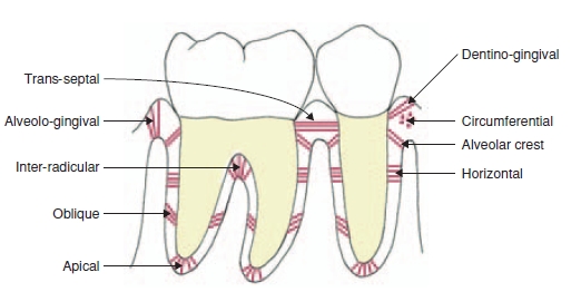
image56 Development and Structure of the Periodontal Ligament

Periodontal Ligament Groups


The periodontal ligament, commonly abbreviated as the PDL, is a group of specialized connective tissue fibers that essentially attach a tooth to the alveolar bone within which it sits.

Features

Functions of PDL are supportive, sensory, nutritive, and remodeling.
The PDL substance has been estimated to be 70% water, which is thought to have a significant effect on the tooth's ability to withstand stress loads. The completeness and vitality of the PDL are essential for the functioning of the tooth.
The PDL ranges in width from 0.15 to0.38 with its thinnest part located in the middle third of the root.
The PDL is a part of the periodontium that provide for the attachment of the teeth to the surrounding alveolar bone by way of the cementum.
The PDL appears as the periodontal space of 0.4 to 1.5 mm on radiographs, a radiolucent area between the radiopaque lamina dura of the alveolar bone proper and the radioopaque cementum.
There are progenitor cells in the periodontal ligament that can differentiate into osteoblasts for the physiological maintenance of alveolar bone and, most likely, for its repair as well.

Structure

The PDL consist of cells, and extracellular compartment of fibers. The cells are fibroblast, epithelial cells, undifferentiated mesenchymal cells, bone and cementum cells. The epithelial rests of Malassez are also present; these groups of epithelial cells become located in the mature PDL after the disintegration of Hertwig epithelial root sheath during the formation of the root. The extracellular compartment consists of collagen fibers bundles embedded in intercellular substance. The PDL collagen fibers are categorized according to their orientation and location along the tooth.

Alveodental ligament

The main principal fiber group is the alveolodental ligament, which consists of five fiber subgroups: alveolar crest, horizontal, oblique, apical, and interradicular on multirooted teeth. Another principal fiber other than the alveolodental ligament are the transseptal fibers.
All these fibers help the tooth withstand the naturally substantial compressive forces which occur during chewing and remain embedded in the bone. The ends of the principal fibers that are within either cementum or alveolar bone proper are considered Sharpey fibers.

Alveolar crest fibers

Alveolar crest fibers (I) extend obliquely from the cementum just beneath the junctional epithelium to the alveolar crest. These fibers prevent the extrusion of the tooth and resist lateral tooth movements.

Horizontal fibers

Horizontal fibers (J) attach to the cementum apical to the alveolar crest fibers and run perpendicularly from the root of the tooth to the alveolar bone..

Oblique fibers

Oblique fibers (K) are the most numerous fibers in the periodontal ligament, running from cementum in an oblique direction to insert into bone coronally.

Apical fibers

Apical fibers are found radiating from cementum around the apex of the root to the bone, forming base of the socket or alveolus.

Interradicular fibers

Interradicular fibers are only found between the roots of multirooted teeth, such as premolars and molars. They also attach from the cementum and insert to the nearby alveolar bone.

Transseptal fibers

Transseptal fibers (H) extend interproximally over the alveolar bone crest and are embedded in the cementum of adjacent teeth; they form an interdental ligament. These fibers keep all the teeth aligned. These fibers may be considered as belonging to the gingival tissue because they do not have an osseous attachment.

Pathology

  • Damage to the PDL may result in ankylosis of the tooth to the jawbone, making the tooth lose its continuous eruption ability. Dental trauma, such as subluxation, may cause tearing of the PDL and pain during function (eating).
  • The epithelial rests of Malassez can become cystic, usually forming nondiagnostic, radiolucent apical lesions that can be seen on radiographs. This occurs as a result of chronic periapical inflammation after pulpitis occurs and must be surgically removed.
  • The PDL also undergoes drastic changes with chronic periodontal disease that involves the deeper structures of the periodontium with periodontitis. The fibers of the PDL become disorganized, and their attachments to either the alveolar bone proper or cementum through Sharpey fibers are lost because of the resorption of these two hard dental tissue.
  • When traumatic forces of occlusion are placed on a tooth, the PDL widens to take the extra forces. Thus, early occlusal trauma can be viewed on radiographs as a widening of the periodontal ligament space. Thickening of the lamina dura in response is also possible. Clinically, occlusal trauma is noted by the late manifestation of increased mobility of the tooth and possibly the presence of pathological tooth migration.

References

  1. Jump up
     Herbert F. Wolf; Klaus H. Rateitschak (2005). Periodontology. Thieme. pp. 12–. ISBN 978-0-86577-902-0. Retrieved 21 June 2011.

  2. Jump up to:
     Illustrated Dental Embryology, Histology, and Anatomy, Bath-Balogh and Fehrenbach, Elsevier, 2011, page 184.
  3. Jump up
     Max A. Listgarten, University of Pennsylvania and Temple University at http://www.dental.pitt.edu/informatics/periohistology/en/gu0401.htm
  4. Jump up
     Ten Cate's Oral Histology, Nanci, Elsevier, 2013, page 220
  5. Jump up to:
     Structure of periodontal tissues in health and disease, ANTONIO NANCI & DIETER D. BOSSHARDT, Periodontology 2000, Vol. 40, 2006, 11–28 athttp://www.nancicalcifiedtissuegroup.com/documents/Nanci%202006.pdf
  6. Jump up
     Max A. Listgarten, University of Pennsylvania and Temple University, http://www.dental.pitt.edu/informatics/periohistology/en/gu0404.htm
  7. Jump up
     Ten Cate's Oral Histolog, Nanci, Elsevier, 2013, page 274
  8. Jump up
     Zadik Y (December 2008). "Algorithm of first-aid management of dental trauma for medics and corpsmen". Dent Traumatol 24 (6): 698–701. doi:10.1111/j.1600-9657.2008.00649.x.PMID 19021668.

Our Mouth -- Diagram

File:Illu mouth.jpg

Understanding our Mouth -- GINGIVA

Gingiva

General description

Gingiva are part of the soft tissue lining of the mouth. They surround the teeth and provide a seal around them. Compared with the soft tissue linings of the lips and cheeks, most of the gingivae are tightly bound to the underlying bone which helps resist the friction of food passing over them. Thus when healthy, it presents an effective barrier to the barrage of periodontal insults to deeper tissue. Healthy gingiva are usually coral pink, but may contain melanin pigmentation.
Changes in color, particularly increased redness, together with edema and an increased tendency to bleed, suggest an inflammation that is possibly due to the accumulation of bacterial plaque. Overall, the clinical appearance of the tissue reflects the underlying histology, both in health and disease. When the gingival tissue is not healthy, it can provide a gateway for periodontal disease to advance into the deeper tissue of the periodontium, leading to a poorer prognosis for long-term retention of the teeth. Both the type of periodontal therapy and homecare instructions given to patients by dental professionals and restorative care are based on the clinical conditions of the tissue.

Macroscopic features of gingiva

The gingiva is divided anatomically into marginal, attached and interdental areas.

Marginal gingiva

The marginal gingiva is the terminal edge of gingiva surrounding the teeth in collar like fashion. In about half of individuals, it is demarcated from the adjacent, attached gingiva by a shallow linear depression, the free gingival groove. This slight depression on the outer surface of the gingiva does not correspond to the depth of the gingival sulcus but instead to the apical border of the junctional epithelium. This outer groove varies in depth according to the area of the oral cavity; the groove is very prominent on mandibular anteriors and premolars.
The marginal gingiva varies in width from 0.5 to 2.0 mm from the free gingival crest to the attached gingiva. The marginal gingiva follows the scalloped pattern established by the contour of the cementoenamel junction (CEJ) of the teeth. The marginal gingiva has amore translucent appearance than the attached gingiva, yet has a simi- lar clinical appearance, including pinkness, dullness, and firmness. In contrast, the marginal gingiva lacks the presence of stippling, and the tissue is mobile or free from the underlying tooth surface, as can be demonstrated with a periodontal probe. The marginal gingiva is stabilized by the gingival fibers that have no bony support. The gingival margin, or free gingival crest, at the most superficial part of the marginal gingiva, is also easily seen clinically, and its location should be recorded on a patient’s chart.

Attached gingiva

The attached gingiva is continuous with the marginal gingiva. It is firm, resilient, and tightly bound to the underlying periosteum of alveolar bone. The facial aspect of the attached gingiva extends to the relatively loose and movable alveolar mucosa, from which it is demarcated by the mucogingival junction. Attached gingiva may present with surface stippling. The tissue when dried is dull, firm, and immobile, with varying amounts of stippling. The width of the attached gingiva varies according to its location. However, certain levels of attached gingiva may be necessary for the stability of the underlying root of the tooth.

Interdental gingiva

The interdental gingiva occupies the gingival embrasure, which is the interproximal space beneath the area of tooth contact. The interdental papilla can be pyramidal or have a "col" shape. Attached gingiva is resistant to masticatory forces and always keratinised.
The col varies in depth and width, depending on the expanse of the contacting tooth surfaces. The epithelium covering the col consists of the marginal gingiva of the adjacent teeth, except that it is nonkeratinized. It is mainly present in the broad interdental gingiva of the posterior teeth, and generally is not present with those interproximal tissue associated with anterior teeth because the latter tissue is narrower. In the absence of contact between adjacent teeth, the attached gingiva extends uninterrupted from the facial to the lingual aspect. The col may be important in the formation of periodontal disease but is visible clinically only when teeth are extracted.

Interdental Areas

It is the part of gingiva which extends in between two teeth up to the contact point.There is a facial side interdental papilla and a lingual side interdental papilla.Interdental papilla has a summit(tip)and margins that are concave.The tip and the margins are unattached and the central portion attached.In inflammations the interdental papilla loses its concavity.

Characteristics of healthy gingiva

Color

Healthy gingiva usually has a color that has been described as "coral pink." Other colours like red, white, and blue can signify inflammation (gingivitis) or pathology. Although described as the colour coral pink, variation in colour is possible. This can be the result of factors such as: thickness and degree of keratinization of the epithelium, blood flow to the gingiva, natural pigmentation, disease and medications.
Since the colour of the gingiva can vary, uniformity of colour is more important than the underlying color itself. Excess deposits of melanin can cause dark spots or patches on the gums (melanin gingival hyperpigmentation), especially at the base of the interdental papillae. Gum depigmentation (aka gum bleaching) is a procedure used in cosmetic dentistry to remove these discolorations.

Contour

Healthy gingiva has a smooth arcuate or scalloped appearance around each tooth. Healthy gingiva fills and fits each interdental space, unlike the swollen gingiva papilla seen in gingivitis or the empty interdental embrasure seen in periodontal disease. Healthy gums hold tight to each tooth in that the gingival surface narrows to "knife-edge" thin at the free gingival margin. On the other hand, inflamed gums have a "puffy" or "rolled" margin.

Texture

Healthy gingiva has a firm texture that is resistant to movement, and the surface texture often exhibits surface stippling. Unhealthy gingiva, on the other hand, is often swollen and less firm. Healthy gingiva has an orange-peel like texture to it due to the stippling.

Reaction to disturbance

Healthy gums usually have no reaction to normal disturbance such as brushing or periodontal probing.
 Unhealthy gums on the other hand will show bleeding on probing (BOP) and/or purulent exudate.

References

  1.  Illustrated Dental Embryology, Histology, and Anatomy, Bath-Balogh and Fehrenbach, Elsevier, 2011, page 123
  2.  Gingival Recession - Causes and treatment JADA, Vol 138. http://jada.ada.or. Oct 2007. American Dental Association
  3.  Mexicodentaldirectory.com > dental sensitivity Retrieved on August 2010
  4.  Mondofacto medical dictionary > gingival retraction 05 Mar 2000
  5.  Mosby's Medical Dictionary, 8th edition. © 2009, Elsevier.